Foraminal Stenosis: Here’s Everything You Need to Know
how we can help
Foraminal Stenosis Overview
Foraminal stenosis is a spinal condition caused by the restriction or abnormal narrowing of the openings within the spine that are known as the foramen.
The medical term for an abnormal narrowing or constriction of the diameter of a bodily passage or orifice is “stenosis.” When the passageway or “canal” that houses the spinal cord and nerve roots is restricted or narrowed, this condition is called spinal stenosis. When the location of the compression is found to be the foramen, the condition is further qualified as foraminal stenosis.
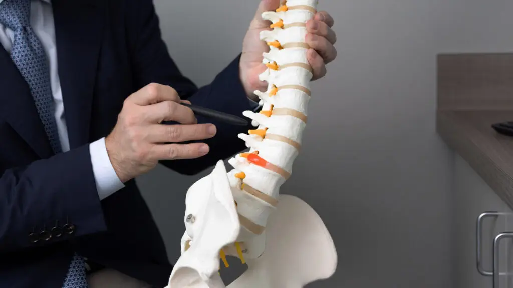
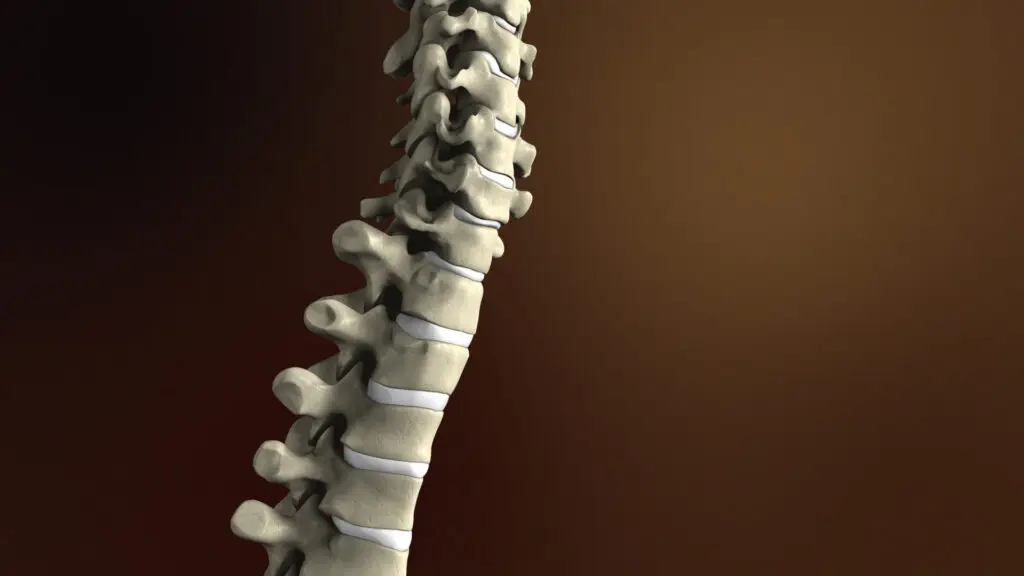
The vertebral foramen is a large opening within the spinal column. Formed by the vertebral body and vertebral arch, it houses and protects the spinal cord, safeguarding it from mechanical damage. Another structure within the spinal column related to the vertebral foramen is the intervertebral foramina. Also known as the intervertebral neural foramen or the intervertebral spinal foramen, spinal nerves exit the spinal column through these openings formed by adjacent vertebrae.
Table of Contents
Symptoms
Diagnosing
Treatments
The Spinal Cord
The spinal cord is a critical component of the nervous system. It functions as a relay station to ensure communication between the brain and the rest of the body. Sensory information like touch, temperature, and pain are transmitted by the spinal cord from peripheral nerves to be processed by the brain. Motor commands from the brain are sent back through the spinal cord to the muscles and internal organs.
The spinal cord controls some of our reflexes without even involving the brain. These involuntary reflexes are called reflex arcs. For example, when you touch something that is hot, the spinal cord initiates an immediate muscle contraction, bypassing the brain’s conscious processing, and sends an immediate signal to move the hand away.
When the spinal cord is compressed, it can lead to various symptoms and functional changes, which may include pain and discomfort, nerve dysfunction, motor and sensory impairments. It may also lead to conditions like sciatica or cauda equina syndrome.

Patients Ask:
What causes Spinal Stenosis?
Texas Back Responds: A variety of factors can lead to spinal stenosis. Disk herniation or bulging discs can cause narrowing in the spinal canal. Facet joints located on the sides of the canal can also contribute to this condition. As the spine ages, the “wear and tear” on the facet joints can lead to what are called bone spurs or cysts which can cause this compression of the nerves. Finally, on the back of the spinal canal, some patients can experience abnormal growth of ligaments which can cause spinal stenosis.
What is Foraminal Stenosis?
Foraminal stenosis is a type of spinal stenosis that occurs at the neural foramen, the openings where spinal nerves exit the spinal column. Foraminal stenosis occurs when the spinal disk between the vertebrae bulges or when the spine joints develop arthritis, causing the foramen to narrow. As a result of this narrowed passageway, the spinal nerve roots that exit the spinal column through the neural foramina may become compressed, leading to pain, numbness, or weakness. When both sides of the foraminal canal narrow, it is referred to as bilateral neural foraminal stenosis.
The development of symptoms from foraminal stenosis will often start gradually and worsen over time. These symptoms can develop on one side or on both sides of the spine. Symptoms may also vary depending on which part of the spine narrows and pinches a spinal nerve.
- Cervical foraminal stenosis occurs in the neural foramens of the neck.
- Thoracic foraminal stenosis occurs in neural foramens in the mid back.
- Lumbar foraminal stenosis develops in the neural foramina of the low back

Lumbar Foraminal Stenosis
The lumbar region, or low back, is the most common area for foraminal stenosis to occur. Depending on where they are in the “spine stack,” vertebrae provide different degrees of load bearing and flexibility for the spine. A combination of evolution and “wear and tear” from aging, work, and play have not been kind to the lumbar region, especially the L4-L5 vertebrae.
The L4-L5 spinal segments are the two lowest vertebrae in the lumbar spine, providing motion in multiple directions and supporting the upper body. These vertebrae undergo heavy impact on daily motion and are highly flexible. Unfortunately, this also means they are highly susceptible to injury and chronic conditions, including spinal stenosis. Lumbar foraminal stenosis is most often the result of age-related spinal degeneration, which can lead to changes in the spine that trigger stenosis.
These changes include:
- Osteoarthritis : Arthritis is the degeneration of any joint in the body and is the most common cause of spinal stenosis.
- Bone Spurs : An outgrowth of extra bone that is usually the result of osteoarthritis or injury. They can form anywhere in the body, but generally appear where bones connect and are most common in the spine.
- Bulging or Herniated Disks : Age-related spinal degeneration can cause the spinal disks to wear down, dry out, and thin. This decompression can put pressure on the spinal nerves or the spinal cord.
- Thickened Spinal Ligaments : Spinal ligaments can thicken or harden over time as part of the aging process, causing a narrowing in the spinal canal that could cause spinal stenosis.
Cervical Foraminal Stenosis
Cervical spinal stenosis occurs when the intervertebral foramina in the cervical spine become narrowed, leading to spinal nerve compression and a range of symptoms like pain and numbness.
As the spinal disks begin to degenerate, they lose height and can bulge out. The disks lose water content, thin out, and become stiffer. This causes the vertebrae to move closer and bone spurs can form around the collapsed disk. Bone spurs contribute to the stiffening of the spine and can cause narrowing of the foramen where the nerve roots exit the spine. This narrowing of the foramen can result in a “pinched nerve.”
The C5-C6 spinal motion segment of the cervical spine provides support and flexibility to the neck and head. Degeneration of the C5-C6 vertebrae and intervertebral disks occur at a higher rate than other cervical vertebrae.
When cervical foraminal stenosis occurs at the C5-C6 vertebrae as the result of spondylosis (degeneration), it usually results in the formation of bone spurs that can cause stenosis or narrowing of the intervertebral foramina. Cervical spinal stenosis can lead to symptoms like pain, numbness, and weakness in the arms, however, not all individuals with cervical spinal stenosis experience symptoms.
The Impact of Uncovertebral Joints
The uncovertebral joints are small synovial joints found between the cervical vertebra from C3-C7. The uncovertebral joints provide flexibility, assist movement, and create stability within the neck and limit sideways movement. Additionally, the uncovertebral joints work to protect the spinal disks and prevent slipped disks.
Another important function of these joints is to protect the intervertebral foramen. Spondylosis or bone spurs in the uncovertebral joints can result in enlargement, or uncovertebral hypertrophy, or enlargement of this tissue.
Some of the symptoms associated with uncovertebral hypertrophy include swelling in the neck area, pain, stiffness, and a grinding or popping noise when moving the neck. Many times, patients report that their pain is worse in the morning after sleeping or after a period of inactivity. Some patients may experience headaches, numbness, or a tingling sensation in their arms, hands, or fingers. It is also possible that a patient will have pain that radiates further down the spine.
Heterotopic ossification is a condition in which bone tissue develops in soft tissues. People with this condition may feel a bony lump under the skin, which can be painful. This condition can also restrict movement.

Patients Ask:
Is Stenosis the Same as a Pinched Nerve?
Texas Back Institute Responds: Stenosis can lead to a pinched nerve, but they are not the same thing. A pinched nerve occurs when pressure is applied to a nerve by a surrounding structure, such as bone, disks, or muscles. A pinched nerve can be the result of stenosis, but also can result from disk herniation, inflammation, or muscle spasm.
Symptoms of Foraminal Stenosis
The spine specialists at Texas Back Institute are experienced in the detection of spinal stenosis. Expertise in diagnosing and treating this condition is fundamental since many of the symptoms can vary depending on where the stenosis is occurring in the spine.
Patients who have stenosis in the neck or cervical region can develop shooting pain in their arms. This can lead to severe pain or chronic pain. They can experience tingling that feels like pins and needles or numbness in the arms. One of the more concerning symptoms of severe stenosis in the neck area is called cervical myelopathy, which is a condition that occurs when the spinal cord at the cervical region becomes compressed.
When this occurs, patients can develop clumsiness with their hands or stumble when walking. They could develop headache symptoms. Treating headaches or migraines is much different than treating cervical stenosis, which is why it is imperative that a trained spine surgeon is consulted for any neck related symptoms.
In the thoracic, or mid-back region, foraminal stenosis will impinge on the spinal cord. This will result in mid back pain. It can also cause leg problems such as numbness or weakness in the legs. This type of stenosis could affect the shoulders and the rib cage. This is the least likely location for this condition.
Spinal stenosis in the low back area can cause symptoms that include shooting pains down the legs, numbness, tingling or weakness in the legs.
If the symptoms of spinal stenosis originate at the L4-L5 vertebrae in the lumbar spine, symptoms could be more pronounced, include:
- Debilitating pain in the lower back and leg
- Sexual dysfunction
- Constant pain, numbness, and/or tingling in the legs when standing
- Bladder or bowel incontinence
- Severe weakness in both legs
- Numbness in the genital and inner thigh area
- “Foot drop,” a condition where it’s impossible to point the ankle and toes upward. This causes the foot to slap onto the ground while walking, with the leg dragging in front of the foot. Foot drops can also throw off balance and cause falls.
Patients Ask:
Is foraminal stenosis of the L4-L5 treated differently than the cervical and thoracic areas?
Texas Back Institute Responds: All foraminal stenosis treatments follow the same protocol, starting with a conservative care approach. If this is not successful, surgery is discussed.
How to Diagnose Nerve Compression from Foraminal Stenosis
One of the critical considerations in the diagnosis of foraminal stenosis involves understanding the problem. The spine specialists at Texas Back Institute begin the process with a diagnostic workup.
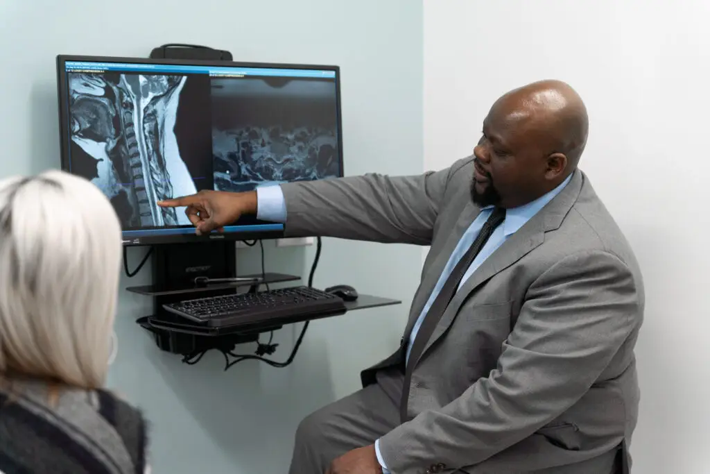
The optimum diagnostic test to detect foraminal stenosis is an MRI (magnetic resonance imaging) scan. This is a medical imaging test that provides physicians with pictures of the spine and the physiological structures inside the body. MRI scanners use powerful magnetic fields, magnetic field gradients, and radio waves to generate images of soft tissues, such as ligaments and disks. MRI scans are particularly useful for evaluating nerve root compression caused by conditions like foraminal stenosis.
An x-ray can be ordered to see the alignment of the spinal vertebrae and the narrowing of the foramen.
If the spine specialists at Texas Back Institute determine that a patient has nerve damage, an electromyography (EMG) may be necessary. An EMG measures muscle response or electrical activity in response to a nerve’s stimulation of the muscle. The test is used to help detect neuromuscular abnormalities by measuring the effectiveness of a patient’s nerves to conduct signals.
A bone scan can be performed to detect arthritis, fractures, infections, and tumors. Bone scans are diagnostic imaging tests that involve using an injection of a radioactive tracer that is carried to the bone that is affected. A special imaging camera picks up the tracer and then sends the information to a computer to create an image that shows bone damage or disease.

Grading the Narrowing of the Foramen
Your spine specialist may grade the level of narrowing of the foramen. The degree of progression will determine treatment options.
- Grade 0 is equal to no foraminal stenosis
- Grade 1 is equal to mild stenosis with no evidence of physical changes to the nerve root
- Grade 2 is equal to moderate stenosis with no evidence of physical changes to the nerve root
- Grade 3 is equal to severe foraminal stenosis showing nerve root collapse
Nerve root compression can occur due to various degenerative causes, such as bone spurs and changes in disc hydration, leading to diagnostic imaging assessments like MRI to evaluate the severity of compression.
Patients Ask:
Are diagnostic procedures such as MRIs, CT myelogram scans, or x-rays safe and effective for patients?
Texas Back Institute Responds: MRI scans, CT scans, and X-rays are all diagnostic tests that allow doctors to see the internal structures of the body. These diagnostic tools create images using various forms of electromagnetic energy. When used by trained technicians, these tools are safe. However, they differ in their accessibility, resolution, and the type of energy they use for imaging.
Treatment for Foraminal Stenosis
Treating foraminal stenosis and the resulting symptoms of nerve compression involves following a protocol that Texas Back Institute has followed since its inception more than 45 years ago. Surgery is ALWAYS the last option to be considered. This is true for every neck and back issue, including foraminal stenosis.
Conservative care is always the first option. In many cases, these conservative care options prove to be highly successful in alleviating a patient’s pain and restoring the patient’s quality of life. These measures include prescribing medication for relief of symptoms and chronic pain, physical therapy, and/or injections.
Foraminal Decompression
In cases where these conservative options prove unsuccessful in alleviating symptoms, surgery is discussed to solve the problem. In the case of spinal stenosis, the primary problem is the narrowing of the spinal canal. The objective of the surgical procedure is to open up that restricted area of the spine and decompress the nerve roots. In many cases, this procedure is as simple as performing a decompression.
With foraminal stenosis, the surgical procedure is called a foraminotomy. Also known as a foraminal decompression, this procedure is performed to relieve pressure on the nerve roots that exit the spinal cord through the intervertebral foramen.
During the procedure, the surgeon will remove a small portion of the bone or tissue to widen the foramen and alleviate the pressure on the nerve and the affected nerve pathway. A foraminotomy can be performed using traditional open surgery or minimally invasive surgical techniques. Recovery from this procedure depends on individual factors and the chosen surgical approach.
Rhizotomy
A minimally invasive procedure called a rhizotomy may be necessary to remove sensation from a painful nerve by killing the nerve fibers responsible for sending pain signals to the brain. This procedure can be used to address different types of pain and abnormal nerve activity. Patients that experience back and neck pain from arthritis, herniated discs, spinal stenosis, and other degenerative spine conditions may be candidates for this procedure.
Spinal Fusion and Artificial Disk Replacement
If the patient has a disk pressing on the spine, the surgeon might recommend replacing it with an artificial disk. This procedure, which was pioneered by the spine surgeons at Texas Back Institute, has proven to be extremely effective in correcting a disk damaged by spinal stenosis.
Other procedures for treating spinal stenosis involve a fusion procedure, where one vertebra is connected, or fused, to another one. This spine surgery is effective when there are concerns about spinal instability.
Patients Ask:
What type of injections are used in the conservative treatment of spinal stenosis?
Texas Back Institute Responds: In most cases, an epidural injection is prescribed for this type of treatment. An epidural involves injecting a medication – either an anesthetic or a steroid – into the space around the spinal nerves known as the epidural space. Steroids help to reduce pain by reducing inflammation around the nerves.

Recovery from Spinal Stenosis Surgery
The advances in diagnostic imaging technology which allow for more accurate location of spinal column restrictions, along with the adoption of minimally invasive surgical procedures, have greatly reduced the time necessary for recovery from spinal stenosis procedures. Most patients return home on the same day as the surgery and are encouraged to begin walking the same day.
Typically, most patients can return to regular movements and life in 4 to 6 weeks after surgery and full results from pain relief might take up to 6 months. During this recovery period, patients recovering from spinal stenosis surgery are recommended to undergo outpatient physical therapy to learn how to move correctly.
Patients Ask:
When should I consult a spine specialist if I am concerned about the symptoms for spinal stenosis?
Texas Back Institute Responds: Once these symptoms become a hindrance to your quality of life, you should see a spine specialist. Pain is a symptom that should not be ignored or be self-treated, particularly if it persists for more than 1-2 weeks. Also, if a patient begins to experience neurological changes such as numbness and weakness or loss of bowel or bladder control, they should seek immediate medical attention.
Lifestyle Adaptations

Many cases of foraminal stenosis are asymptomatic (without symptoms) and patients might not even know that they have this condition, even when there is significant foraminal narrowing. Symptoms usually start out slowly over time and may even seem to come and go.
Foraminal stenosis is most common in people over 50, as it can frequently be associated with degenerative disk disease. When symptoms do develop, many people report that they gradually worsen over time. Symptoms can vary depending on where the stenosis occurs in the spine.
People can make lifestyle changes that will help prevent the onset of spinal stenosis.
These lifestyle adaptations may include:
Maintaining a healthy weight
When a person is overweight, it puts tension on the spine, particularly in the low back, or lumbar region, which is responsible for supporting the weight of the vast majority of the body. Individuals who are overweight also tend to be more inactive which causes their core muscles to weaken. This can also contribute to degenerative spinal conditions because the weakened muscles are not able to provide added support to the spine.
Smoking Cessation
Smoking causes injury and inflammation to the blood vessels, decreasing blood flow to all tissues. This decrease in circulation doesn’t just affect the major arteries, but also causes an impact to the blood supplied to the intervertebral disks of the spine, which are fundamental to spinal stability and proper alignment. Smoking also deprives spinal disks of essential nutrients and interferes with bone metabolism, increasing the risk of spinal fractures.
Exercise
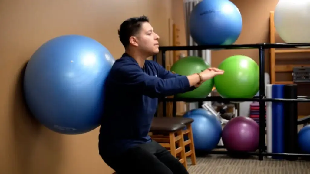
Strong core muscles can help to prevent and treat spinal stenosis. A recent study found that older people with spinal stenosis had better treatment outcomes when they practiced core stability exercises instead of conventional exercises. Some of the core stability exercises included planks and modified push-ups.
Proper Posture
Attention to posture, especially in a seated position, can help to maintain a healthy spine. Researchers suggest that sitting straighter reduces the load on the cervicothoracic joint, the joint that connects the skull to the spine. This is also true of the joints in the lower back.
Avoid “Text Neck”
Spending hours each day staring down at a laptop or mobile phone can be hazardous to your health. Text Neck (link to blog post Text Neck) is defined as an “overuse syndrome” involving the head, neck, and shoulders, usually resulting from excessive strain on the spine from looking in a downward position at hand-held devices, such as smartphones, gaming devices, e-readers, and computer tablets. The most effective tactic to avoid text neck is to reduce the amount of time spent doing this type of activity. It has been estimated that people younger than 30 will send 3,000 texts per month and it is almost impossible to stop this activity cold turkey. However, scheduling “no phone” times during the day can help reduce the amount of time spent texting and still allow someone to conduct business.
Develop Healthy Sleep Habits
The spine specialists at Texas Back Institute (link to blog post back pain and sleep) recommend sleeping on your side or on your back with a pillow beneath your knees. If sleeping on your stomach is the only comfortable position for you, place a pillow under your pelvis or lower abdomen for support. This helps to take pressure off your back.
What’s Next?
If foraminal stenosis, causing nerve compression and other related symptoms, is ruining your quality of life, there’s good news. It doesn’t have to.
Using state-of-the-art medical technology such as robotics, minimally invasive surgery, artificial disk replacement and other tools, the world-class surgeons at Texas Back Institute have successfully diagnosed and treated thousands of patients suffering from the chronic pain of this debilitating condition.
Don’t let foraminal stenosis “impinge” on your quality of life! When you’re ready to become pain-free, click here and get your life back!
Learn more
Frequently Asked Questions
A variety of factors can lead to spinal stenosis. Disk herniation or bulging discs can cause narrowing in the spinal canal. Facet joints located on the sides of the canal can also contribute to this condition. As the spine ages, the “wear and tear” on the facet joints can lead to what are called bone spurs or cysts which can cause this compression of the nerves. Finally, on the back of the spinal canal, some patients can experience abnormal growth of ligaments which can cause spinal stenosis.
Stenosis can lead to a pinched nerve, but they are not the same thing. A pinched nerve occurs when pressure is applied to a nerve by a surrounding structure, such as bone, disks, or muscles. A pinched nerve can be the result of stenosis, but also can result from disk herniation, inflammation, or muscle spasm.
All foraminal stenosis treatments follow the same protocol, starting with a conservative care approach. If this is not successful, surgery is discussed.
MRI scans, CT scans, and X-rays are all diagnostic tests that allow doctors to see the internal structures of the body. These diagnostic tools create images using various forms of electromagnetic energy. When used by trained technicians, these tools are safe. However, they differ in their accessibility, resolution, and the type of energy they use for imaging.
In most cases, an epidural injection is prescribed for this type of treatment. An epidural involves injecting a medication – either an anesthetic or a steroid – into the space around the spinal nerves known as the epidural space. Steroids help to reduce pain by reducing inflammation around the nerves.
Once these symptoms become a hindrance to your quality of life, you should see a spine specialist. Pain is a symptom that should not be ignored or be self-treated, particularly if it persists for more than 1-2 weeks. Also, if a patient begins to experience neurological changes such as numbness and weakness or loss of bowel or bladder control, they should seek immediate medical attention.
Locations


