Cervical Disc Replacement Surgery: Like New!
how we can help
Cervical Disc Replacement Overview
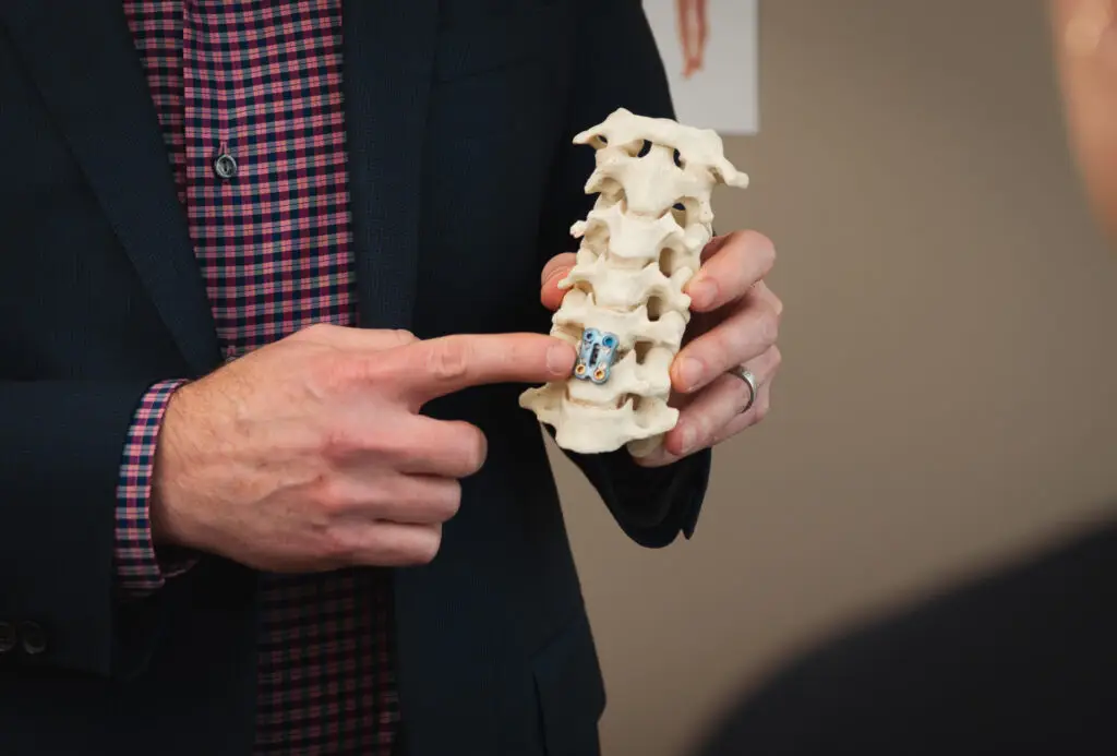
Cervical disc replacement surgery is a procedure to remove a diseased cervical disc and replace it with an implanted artificial disc that can mimic the function of the native disc. It’s designed to preserve and maintain the natural motion of the spine while remaining secured to the surrounding vertebral bones.
Total disc replacement, or TDR, is performed less often than traditional spinal decompression and fusion surgeries. As more outcome studies demonstrate the effectiveness of artificial discs in treating neck and back pain, TDR will become even more widespread.
Pioneers in Artificial Disc Replacement Surgery
Texas Back Institute (TBI) is a pioneer in artificial disc replacement, performing the first procedure in March 2000. As a leader in spinal research and innovation, Texas Back has participated in more than 14 different FDA trials of both cervical and lumbar discs. Alleviating the pain caused by injured or diseased discs has been life-changing for TBI patients. Plus, these procedures and patient follow-ups have provided an extensive trove of data on the efficacy of this ground-breaking treatment.
Table of Contents
Treats
Pain caused by injured or diseased discs. When the space between spinal vertebrae becomes too narrow, part of the vertebrae or the cervical disc can press on the spinal cord or nerves, causing pain, numbness, weakness, or even headaches in some patients. Other conditions, like herniated discs, and “radicular syndromes” are also potential causes.
Diagnosing
Tests and imaging studies to diagnose cervical disc degeneration may include:
Recovery
Patients Ask:
What causes a cervical disk to become diseased?
Texas Back Institute Responds: The most common condition is degenerative disk disease, which leads to the erosion of the vertebral disks in the cervical spine (neck)This typically occurs as people age. Although not actually a “disease,” disk degeneration can be painful and incapacitating. When the spinal discs, which act as shock absorbers cushioning the vertebrae, gradually wear down, the bones can start to rub together. Also, when the space between spinal vertebrae becomes too narrow, part of the vertebrae or the cervical disc can press on the spinal cord or nerves, causing pain, numbness, weakness, or even headaches in some patients. Other conditions, like herniated discs, and “radicular syndromes” are also potential causes. If symptoms don’t respond to conservative care treatments, then cervical artificial disc replacement surgery may be recommended.
Biomechanics of the Cervical Spine
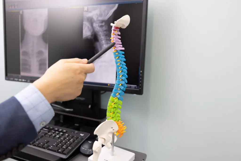
The cervical spine (neck) consists of 7 vertebrae, C1-C7, that connects the skull to the thoracic (mid) spine, allowing the head to move while maintaining stability and protecting the spinal cord.
The first two vertebrae, C1 and C2, are distinct in shape and function, and do not have a disc between them.
- C1: The first vertebra is a ring-shaped bone that begins at the base of the skull. Called the “atlas,” it is named after Atlas of Greek mythology, who held the world on his shoulders. The C1 holds the head in an upright position.
- C2: The second vertebra is called the “axis.” The atlas can pivot against the C2 axis vertebra, which permits the lateral side-to-side rotation of the head, as in when shaking the head “no.”
C1-C7 vertebrae are connected at the back of the bone by the facet joints, which allow the forward, backward, and twisting movement of the neck. In addition, the cervical vertebrae are surrounded by nerves, muscle, ligaments, and tendons.
Intervertebral discs are positioned between each vertebra. A total of five discs are positioned between the lower six cervical vertebrae (one between two vertebrae). In addition to cushioning against stresses placed on the neck, the discs allow the head to flex and rotate during activity.
The spinal cord travels from the base of the brain to the upper lumbar spine. It sends messages from the brain to the muscles and brings back messages from the sensory receptors. The proper functioning of the spinal cord is required for movement of the arms and legs as well as for receiving sensations such as touch, pain and temperature.

Patients Ask:
What are the benefits of Cervical Disc Replacement?
Texas Back Institute Responds: Cervical disc replacement has many benefits, first and foremost, is alleviating the pain caused by the diseased disc. Other benefits include preserving the motion of the neck and decreasing the chance of future surgery. Spinal fusion surgery can lead to a condition known as “adjacent segment disease,” or ASD, which can be attributed to increased stress at the levels above and below the fusion. ASD can be the result of continuing degenerative disc disease as well.

Cervical Disc Replacement vs. Fusion
There are many factors to take into consideration when deciding whether a patient is a good candidate for cervical disc replacement surgery. Depending on the individual patient, anterior cervical discectomy with fusion, or ACDF, might be the best option. Both procedures have favorable clinical outcomes. As with any surgery, patients should discuss the potential benefits and risks with their surgeon.
ACDF is the most common surgery for treating symptoms related to a degenerative or herniated disc in the neck. This procedure consists of removing the diseased disc entirely and replacing it with a bone graft (or bone graft substitute) to allow the adjacent vertebrae to eventually fuse together. ACDF is still considered the “gold standard” for treating cervical degenerative disc disease because it has been well studied and can treat a greater variety of spinal conditions. It should be noted that many patients have no complications with the bone graft forming a fusion or any issues with adjacent segment disease.
Cervical artificial disc replacement (ADR) is different from ACDF because rather than fusing the adjacent vertebrae together, an artificial disc is inserted to maintain motion between the vertebrae. This allows a more normal biomechanics within the cervical spine. In addition, Cervical ADR places less stress on the adjacent spine segments than ACDF. Unlike ACDF, no bone graft is necessary. In terms of recovery, patients who have cervical ADR are cleared for more strenuous activities in about 6 weeks, as opposed to the 3 months that’s typical for ACDF.
Patients Ask:
Are there any conditions that would disqualify a patient from receiving a cervical disc replacement?
Texas Back Institute Responds: Cervical artificial disc replacement surgery is not recommended for patients with advanced spinal degeneration from ossified posterior longitudinal ligament, a condition in which a flexible structure known as the posterior longitudinal ligament becomes thicker and less flexible, or degenerating facet joints associated with osteoarthritis or ankylosing spondylitis. Patients with weakened bones, like those with osteoporosis, are generally not good candidates for cervical ADR. Other considerations are a prior cervical surgery that may have altered the anatomy or patients with an allergic reaction to metal that may disqualify a candidate.
How is Degenerative Disc Disease Diagnosed?
First, spine specialists will take a thorough medical history, ask about your symptoms, perform a physical exam, and order tests and imaging studies.
Tests and imaging studies to diagnose cervical disc degeneration may include:
- Computed tomography (CT) scan: This is a scan that uses X-rays and computers to produce images that are “slices” of the area under examination. A CT scan will show an image of the shape and size of the spinal canal, its contents and the bone around it. It helps diagnose bone spurs, osteophytes (same as bone spurs), arthritis, bone fusion and bone destruction from infection or tumor.
- Magnetic resonance imaging (MRI): Using a large magnet, radio waves and a computer, MRIs produce detailed images. This type of scan can show problems with the spinal cord and the nerves exiting the spinal column, spinal degeneration, disc herniation, and more.
- X-rays: X-rays create pictures of the bones and soft tissues, using a small amount of radiation. X-rays can show fractures, disc problems, spinal alignment problems and the presence of arthritis.
- Electromyogram (EMG) and nerve conduction studies: An EMG helps evaluate the health and function of nerves and muscles. A nerve conduction study measures how fast an electrical impulse moves through the nerves. These tests help determine ongoing nerve damage and the site of nerve compression.
- Myelogram: This imaging test examines the relationship between vertebrae and discs and outlines the spinal cord and nerves exiting the spinal column. It shows possible things such as a tumor, bone spurs or herniated disc that are pressing against the spinal cord, nerves or nerve roots and causing pain, numbness or weakness.
Patients Ask:
If my surgeon thinks I am a good candidate, what should I expect?
Texas Back Institute Responds: Cervical arthroplasty is a relatively safe procedure. Surgeons make an incision on the front of the neck, just off of midline – which is relatively pain free since only a small incision is necessary. The trachea and esophagus are moved to the side and blood vessels are moved allowing the diseased disc to be removed using magnification and ensuring that the spinal cord and all the nerves are completely decompressed.
The artificial disc is then implanted using an intraoperative x-ray so that it’s positioned appropriately. The incision is closed with dissolvable sutures. Patients may be given a soft collar to wear to help protect the wound. Some patients stay at the hospital overnight, especially if they are having multiple levels done, but many times patients can go home the same day.
Recovery From This Procedure
Recovery time is typically shorter with cervical ADR because the vertebrae don’t need to fuse together. Recovery and rehabilitation at home may be a little different for each person, but in general, here’s what to expect:
- Patients may need to continue to wear a soft collar
- Ask about activity and bathing restrictions
- Expect to make a gradual return to normal activity
- Expect to start physical therapy after a few weeks
- A return to full activity within 4-6 weeks
Patients should follow their prescribed care following any cervical spine surgery, which may include rest, pain management, and some restrictions. Most people experience significant improvement in pain during the second week after surgery and little to no pain after they heal. A return to driving and work depends on the recovery process and type of work.

Patients Ask:
What is the success rate of cervical disc replacement?
Texas Back Institute Responds: TBI has been at the forefront in artificial disc replacement, which allowed us to gain valuable clinical data about the success rate of our patients. In fact, success rates are about 90% across clinical studies, and patient satisfaction scores are remarkably high. The best chance of successful recovery comes from choosing a spine specialist, like those at TBI, with extensive experience performing artificial disc replacement surgery.
Why Wait?
Improved materials, technology and surgical advancements in artificial disc replacement have revolutionized treatment for this crucial component of the spine. Since the first tests, Texas Back Institute has been one of the world-wide leaders in this procedure. If you are suffering from neck or back pain, contact the experts at Texas Back Institute today.
Learn more
Frequently Asked Questions
The most common condition is degenerative disk disease, which leads to the erosion of the vertebral disks in the cervical spine (neck)This typically occurs as people age. Although not actually a “disease,” disk degeneration can be painful and incapacitating. When the spinal discs, which act as shock absorbers cushioning the vertebrae, gradually wear down, the bones can start to rub together. Also, when the space between spinal vertebrae becomes too narrow, part of the vertebrae or the cervical disc can press on the spinal cord or nerves, causing pain, numbness, weakness, or even headaches in some patients. Other conditions, like herniated discs, and “radicular syndromes” are also potential causes. If symptoms don’t respond to conservative care treatments, then cervical artificial disc replacement surgery may be recommended.
Cervical disc replacement has many benefits, first and foremost, is alleviating the pain caused by the diseased disc. Other benefits include preserving the motion of the neck and decreasing the chance of future surgery. Spinal fusion surgery can lead to a condition known as “adjacent segment disease,” or ASD, which can be attributed to increased stress at the levels above and below the fusion. ASD can be the result of continuing degenerative disc disease as well.
Cervical artificial disc replacement surgery is not recommended for patients with advanced spinal degeneration from ossified posterior longitudinal ligament, a condition in which a flexible structure known as the posterior longitudinal ligament becomes thicker and less flexible, or degenerating facet joints associated with osteoarthritis or ankylosing spondylitis. Patients with weakened bones, like those with osteoporosis, are generally not good candidates for cervical ADR. Other considerations are a prior cervical surgery that may have altered the anatomy or patients with an allergic reaction to metal that may disqualify a candidate.
Cervical arthroplasty is a relatively safe procedure. Surgeons make an incision on the front of the neck, just off of midline – which is relatively pain free since only a small incision is necessary. The trachea and esophagus are moved to the side and blood vessels are moved allowing the diseased disc to be removed using magnification and ensuring that the spinal cord and all the nerves are completely decompressed.
The artificial disc is then implanted using an intraoperative x-ray so that it’s positioned appropriately. The incision is closed with dissolvable sutures. Patients may be given a soft collar to wear to help protect the wound. Some patients stay at the hospital overnight, especially if they are having multiple levels done, but many times patients can go home the same day.
TBI has been at the forefront in artificial disc replacement, which allowed us to gain valuable clinical data about the success rate of our patients. In fact, success rates are about 90% across clinical studies, and patient satisfaction scores are remarkably high. The best chance of successful recovery comes from choosing a spine specialist, like those at TBI, with extensive experience performing artificial disc replacement surgery.
Locations


