Getting to the Root of the Problem with Decompression Surgery
how we can help
Decompression Surgery Overview
One of the most common reasons people miss work each year is from back pain. It’s the leading cause of disability worldwide. According to the American Association of Neurological Surgeons, “75 to 85 percent of Americans will experience back pain in their lifetime. Of those, 50 percent will have more than one episode within a year.”
In some cases, decompression surgery or other forms of spinal decompression are used to alleviate this back pain. Decompression surgery is any surgery that relieves compression of the spinal cord or the spinal nerves by creating space around the affected area. Although decompression procedures vary, the desired outcome remains the same.
Our spinal cord contains 31 pairs of nerves running the length of the spinal column. These nerve roots branch off through small openings in the vertebrae. Spinal nerves are responsible for providing movement and sensation to different areas of the body. Nerve compression, sometimes called a pinched spinal nerve, can occur anywhere along the spine, from the top (cervical spine) to the bottom (sacrum).
The lumbar spine is particularly susceptible to nerve compression, often necessitating decompression surgery to relieve symptoms.
Table of Contents
Treats
Diagnosing
Recovery
Conditions that can Cause Spinal Compression
Many conditions can cause compression of spinal cord and the nerve roots, or spinal stenosis. Stenosis is defined as pressure on the nerves of the spine. For example, a disc bulge or herniation can take up space for the nerves. Bone spurs and overgrown, soft tissues, the result of arthritis, can cause pressure on the spinal cord. Age related “wear and tear” problems can also take up space, causing nerves to be compressed. Spinal stenosis is most common in the lower (lumbar region) part of the spine and upper (cervical region) part of the spine, producing similar symptoms, but in different parts of the body.
A Brief Look at the History of Decompression Surgery
The 1970’s transformed the ability to diagnose complex issues in the spine. Doctors were able to visualize spinal structures using diagnostic imaging like spinal computerized tomography, (CT scan), and magnetic resonance imaging, (MRI). In 1975, the first procedure to remove a damaged disk through the skin was performed. New instruments were being introduced to better protect the spinal cord during surgery. By monitoring the functioning of the nerve roots and alerting the surgeon with an early warning signal, these new instruments helped prevent damage to the spinal cord. While screws or plates were being set, the surgeon could be confident that the spinal cord was protected.
In the 1980’s, discectomy procedures continued to improve using new instruments and smaller incisions. By 1985, disk decompression was accomplished by the partial removal of disk material through the skin. Instruments initially used for delicate eye surgeries were redesigned for minimally invasive spine procedures.
In the hands of a skilled surgeon, technology can become a powerful force for healing. Texas Back Institute has become a world leader in endoscopic spine surgery, and hundreds of patients obtain life-changing relief from pain every year.
In 2000, Texas Back Institute brought artificial disc replacement (ADR) technology to the United States and performed the first ADR procedure. TBI surgeons were the first to perform lumbar disc replacement in the US and have published important research papers on cervical and lumbar arthroplasty.
Advancements in spinal decompression surgeries, including the use of various types of anesthesia and minimally invasive techniques, have significantly improved patient outcomes.

(illustration of spine with stenosis)
Symptoms of Stenosis
People can have stenosis and experience no symptoms, but if symptoms begin to manifest, treatment is needed. Nerve compression symptoms are often present in the arms or legs, not just in the neck or back. These syptoms can present themselves as arm pain, leg pain, as well as neck or back pain. If a patient experiences pain, numbness, tingling, or muscle weakness in their extremities, then stenosis or nerve compression is most likely causing the symptoms.
If spinal stenosis patients report a feeling of heaviness, discomfort, achiness, and generalized weakness in their buttocks and legs that becomes worse when they stand and walk and better when they sit and lean forward, the lumbar region of the spine is affected. So, even before looking at an MRI, spine specialists can detect the symptoms of stenosis in the lower back.
Patients Ask:
What tests are used to diagnose stenosis?
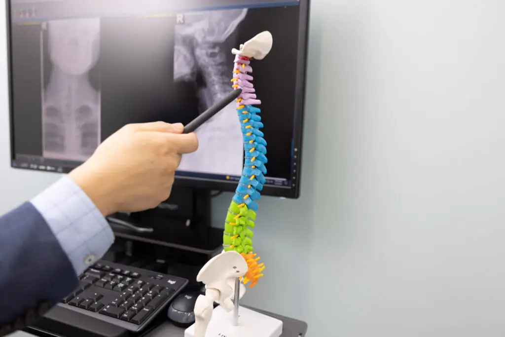
Texas Back Institute Responds: Spinal stenosis can be diagnosed using several different modalities. Diagnosis begins by taking a medical history and talking to a patient about the symptoms they are experiencing. A physical examination is performed, and imaging studies are ordered. X-rays that show the structural spine and how a patient stands and moves are a helpful initial step. An MRI will show doctors nerves, soft tissues, and the discs themselves. If a patient is not responding to the early phase of non-surgical treatment, an MRI can help doctors to diagnose exactly the location where the symptoms are coming from and can target the affected area with epidural injections or potentially decompression surgery.
If there are any questions remaining, a doctor may order another ancillary test, like electromyography, or EMG, that might be useful in pinning down the exact location of the stenosis. This test records muscle and nerve activity that might detect abnormalities that are not physically obvious.

Top Two Decompression Surgeries: Laminectomy and Discectomy
Spinal decompression surgery is most often either a laminectomy or a discectomy. A laminectomy addresses the wear and tear on the structure of the spine. Decompression for a bulging disc is called a discectomy, or a microdiscectomy. Although both are decompression surgeries, there is a difference.
A laminectomy involves removing a portion of the vertebral bone called the lamina to relieve pressure on the spinal cord or nerves. A discectomy involves the partial or total removal of the compressed disc that’s causing the symptoms.
Decompression surgery can be done from the back using an open incision. Depending on what needs to be decompressed or cleaned out, and at what levels and how extensive the work is, incision size can vary. Decompression surgery involves opening the back and moving the muscles that overlay the spine so the surgeon can see the spine. The surgeon uses a variety of instruments and tools to take the pressure off – whether it comes from bone spurs, disc bulge, or some combination of both. Patients wake up from surgery and immediately report incredible improvement in their arm or leg symptoms.
Less invasive decompression surgery can be performed using a tubular retractor. The surgeon operates through a tube using special magnifying lenses or a microscope. This allows the surgeon to do a similar procedure with a smaller incision and much less muscle disruption, which allows for a faster recovery.
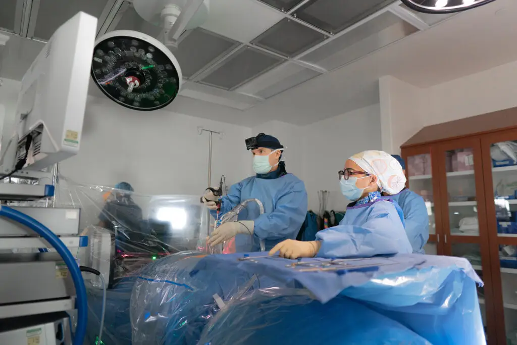
At the least invasive end of the spectrum, there are endoscopic techniques that can accomplish the exact goals of decompression surgery, with an even smaller incision and even less muscle and soft tissue disruption. Patients may even go home the same day, depending on how the decompression surgery is performed.
Other types of decompression surgery include:
- Corpectomy: Removal of a vertebra and the disks attached above and below it. An artificial implant fills the remaining space.
- Foraminotomy: Widens the foramen, the opening where a nerve root leaves the spinal canal.
- Laminoplasty: Opening the lamina rather than removing it by creating a hinge in the bone, allowing more room in the spinal canal.
- Laminotomy: Like a laminectomy, removing only a small portion of the bone.
- Osteophyte Removal: Osteophytes, or bone spurs, can cause pressure on spinal nerves.
- Anterior Lumbar Interbody Fusion (ALIF): Surgeons enter through the abdomen to reach the front of the spine to remove and replace an intervertebral disk with bone or a metal spacer.
Patients Ask:
Will I need decompression surgery?
Texas Back Institute Responds: At Texas Back Institute, surgery is always a last resort. Our doctors initially offer conservative treatment and nonsurgical options like physical therapy, judicious use of non-opioid medications, and epidural injections. In many cases, that’s enough to help patients recover from stenosis symptoms without the need for surgery. However, if symptoms persist, or the patient develops other symptoms that might be considered red flags – like weakness – decompression surgery may be recommended.
Decompression With Stabilization
In some instances, decompression surgery alone is insufficient. The surgeon may need to perform a stabilization in addition to the decompression surgery – not for a bone spur or disc herniation – but because the vertebrae themselves are unstable and moving. This is called spondylolisthesis. Decompression surgery might also require stabilization if the spine has a deformity, or curvature that could be a primary contributor to nerve compression. In cases like this, stabilization to put the bones back where they belong and hold them there is necessary to alleviate pressure from the nerves. This stabilization surgery is called a spinal fusion operation. There are a variety of ways to perform spinal fusion, from the traditional methods to minimally invasive procedures. In some cases, a fusion can be added to decompression surgery if additional stability is needed.
Patients Ask:
What is the recovery period for decompression surgery?
Texas Back Institute Responds: Recovery periods are based on the extensiveness of the surgery and the number of levels impacted. For example, if surgery involves removing bone spurs or disc herniation without the need for a fusion operation or metal placement, a patient could stay in the hospital a day or two, or even go home the same day. Overall, recovery is much faster and smoother for decompression surgery than for other types of fusion or back surgery.
After Decompression Surgery: Next Steps
Intermediate recovery typically encompasses the period from 2 weeks to 6 weeks after surgery. If your surgeon prescribes specific exercises to do after spine surgery, follow their instructions carefully. Certain exercises may be more effective for a faster recovery based on the type of procedure you had. For example, core exercises and hamstring stretches are often recommended after a microdiscectomy, but may be restricted following lumbar fusion surgeries.
Walking several times a day for progressively increasing distances is the single safest and most effective exercise recommendation. Your surgeon may also recommend during this period that you begin to see a physical therapist who will work with you to design a safe, effective exercise program to restore normal daily function.

Patients Ask:
How long does it take to fully recover after back surgery?
Texas Back Institute Responds: Recovery times will vary depending on individual factors regarding your procedure, general medical condition, and patient cooperation. Most patients need six weeks to six months to make a full recovery from decompression surgery. Patients can usually resume light work and drive a car after 2 weeks. Low impact exercise can resume after 4 weeks. More strenuous activity can resume from 8 to 12 weeks.
Why Wait?
If back pain is interfering with your quality of life, it might be time to talk to an expert at Texas Back Institute and set an appointment.
Learn more
Frequently Asked Questions
Spinal stenosis can be diagnosed using several different modalities. Diagnosis begins by taking a medical history and talking to a patient about the symptoms they are experiencing. A physical examination is performed, and imaging studies are ordered. X-rays that show the structural spine and how a patient stands and moves are a helpful initial step. An MRI will show doctors nerves, soft tissues, and the discs themselves. If a patient is not responding to the early phase of non-surgical treatment, an MRI can help doctors to diagnose exactly the location where the symptoms are coming from and can target the affected area with epidural injections or potentially decompression surgery.
If there are any questions remaining, a doctor may order another ancillary test, like electromyography, or EMG, that might be useful in pinning down the exact location of the stenosis. This test records muscle and nerve activity that might detect abnormalities that are not physically obvious.
At Texas Back Institute, surgery is always a last resort. Our doctors initially offer conservative treatment and nonsurgical options like physical therapy, judicious use of non-opioid medications, and epidural injections. In many cases, that’s enough to help patients recover from stenosis symptoms without the need for surgery. However, if symptoms persist, or the patient develops other symptoms that might be considered red flags – like weakness – decompression surgery may be recommended.
Recovery periods are based on the extensiveness of the surgery and the number of levels impacted. For example, if surgery involves removing bone spurs or disc herniation without the need for a fusion operation or metal placement, a patient could stay in the hospital a day or two, or even go home the same day. Overall, recovery is much faster and smoother for decompression surgery than for other types of fusion or back surgery.
Recovery times will vary depending on individual factors regarding your procedure, general medical condition, and patient cooperation. Most patients need six weeks to six months to make a full recovery from decompression surgery. Patients can usually resume light work and drive a car after 2 weeks. Low impact exercise can resume after 4 weeks. More strenuous activity can resume from 8 to 12 weeks.
The artificial disc is then implanted using an intraoperative x-ray so that it’s positioned appropriately. The incision is closed with dissolvable sutures. Patients may be given a soft collar to wear to help protect the wound. Some patients stay at the hospital overnight, especially if they are having multiple levels done, but many times patients can go home the same day.
Locations


