Diagnostic Testing at Texas Back Institute
how we can help
The spine specialists at the Texas Back Institute have an arsenal of diagnostic tests to ensure precise diagnosis and successful treatment.
Table of Contents
These diagnostic tests include:
Discography
A discography, also called a discogram, uses x-rays and a radiographic contrast dye to detect damaged disks. The contrast can reveal any cracks in the disk’s exterior and assess its condition.
Computerized Tomography (CT)
Computed tomography of the spine is a diagnostic imaging test used to help diagnose, or rule out, spinal column damage in injured patients. CT scanning is fast, painless, noninvasive and accurate. Lumbar -spine CT scans evaluate the causes of lower back pain. A CT with myelogram diagnoses conditions like stenosis. This test is prescribed by a physician.
Magnetic Resonance Imaging (MRI)
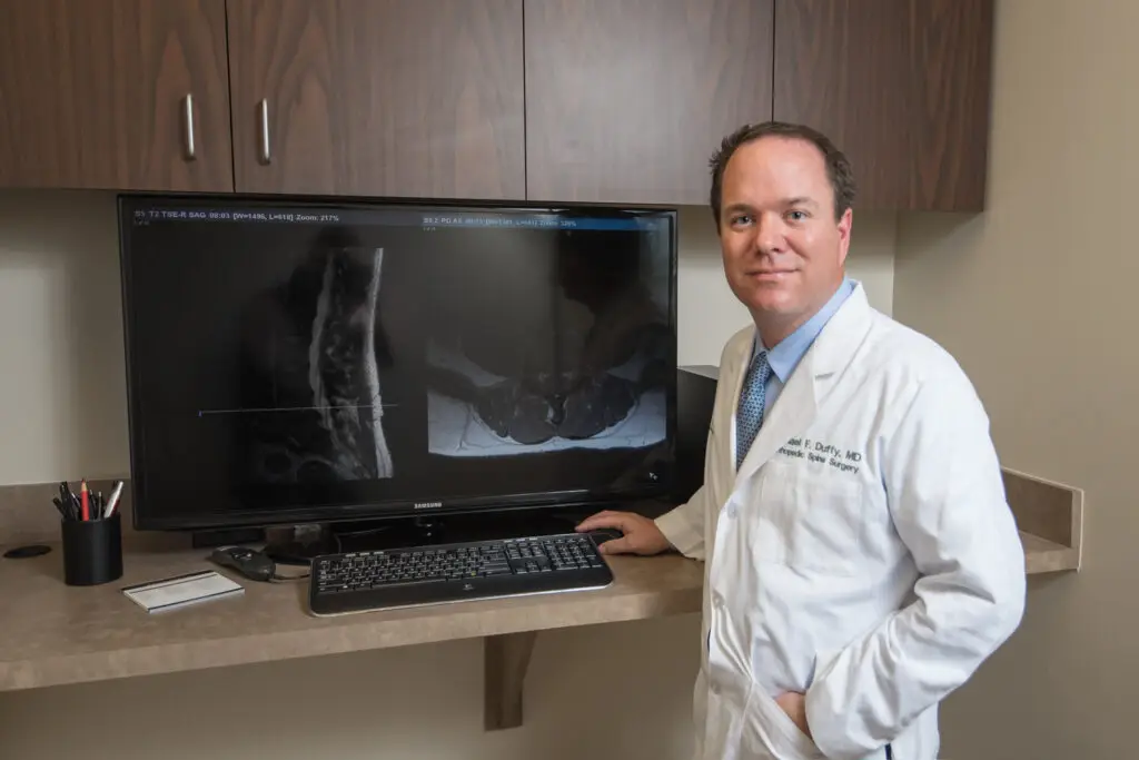
Magnetic resonance imaging of the spine uses radio waves, a magnetic field and a computer to create clear, detailed pictures of the spine and surrounding tissues. Cervical MRIs evaluate the neck region. Thoracic MRIs evaluate the mid-back. Lumbar MRIs evaluate the low back. This test is prescribed by a physician.

X-ray
An X-ray is a quick, painless test that captures images of structures inside the body, particularly the bones. X-ray beams pass through the body, and dense materials like bone and metal appear as white on the X-ray. A cervical spine X-ray focuses on the neck. A thoracic spine X-ray focuses on the mid-back. A lumbar spine X-ray examines the lower back. A coccyx X-ray targets the tailbone. This test is prescribed by a physician.
Myelography
Myelography, or myelogram, is an imaging exam that introduces a needle into the fluid-filled space around the spinal cord and injects a tiny amount of contrast material into this space, called the “subarachnoid space”. A real-time form of x-ray called fluoroscopy allows the radiologist to take moving pictures that evaluate the spinal cord, nerve roots and the membranes that cover them, known as the meninges.

Physical Examination
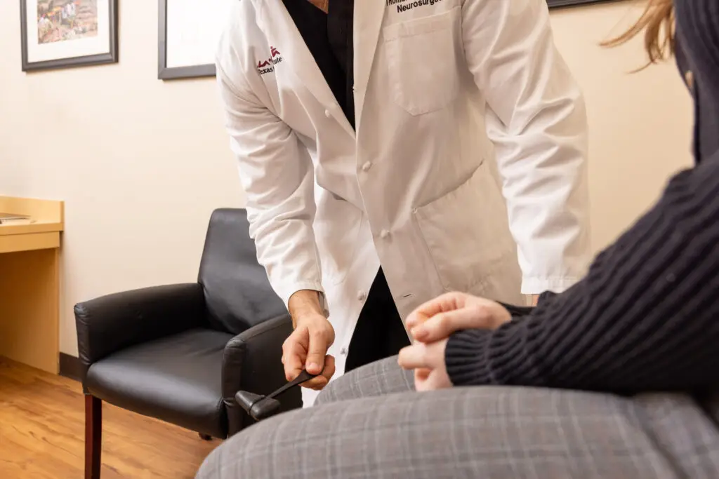
A physical examination continues the diagnostic process, adding information obtained by inspection, palpation, percussion, and auscultation. When data accumulated from the history and physical examination are complete, a working diagnosis is established, and diagnostic tests are selected that will help to retain or exclude that diagnosis.
Injections
Spinal injections are performed under X-ray guidance, called fluoroscopy, to confirm correct placement of the medication and improve safety. Injections can be used in two ways. They can be used for diagnostics and for therapeutic treatment.
Epidural Steroid Injections
Epidural steroid injections (ESIs) are commonly used to manage chronic low back pain caused by spinal conditions. They can also serve as a diagnostic tool to pinpoint the source of pain. A Selective Nerve Root Block targets specific spinal nerves to assess for pain.
Facet Joint Injections
Facet joint injections serve both diagnostic and therapeutic purposes. They help identify the source of facet joint pain while providing pain relief. The injection contains steroids to reduce inflammation and local anesthetics for immediate relief.

Selective Nerve Root Block
A selective nerve root block (SNRB) involves precise administration of a local anesthetic or a combination of anesthetic and steroid around a spinal nerve. Its main purposes are to provide pain relief by reducing inflammation and to help diagnose the specific spinal nerve root causing symptoms.
Nerve Conduction Study
A nerve conduction study (NCS) is a diagnostic test that evaluates the function of your peripheral nerves. This test is often done in conjunction with Electromyography (EMG) to measure electrical current in motor nerves and sensory nerves, to assess for nerve compression syndromes, such as sciatica.
Electromyography
Electromyography (EMG) is a diagnostic procedure that evaluates the health of muscles and the nerve cells controlling them (motor neurons).
Locations


