Rhizotomy: Immediate Relief for Facet and Sacroiliac Joint Pain
how we can help
Rhizotomy Overview
Facet and sacroiliac joint conditions can be very painful. If the pain does not subside with chiropractic care, physical therapy, medication, and other non-invasive treatments, injections may be considered. Depending on the response to these injections, a rhizotomy may be considered.
Rhizotomy, which is also called ablation or neurotomy, is a minimally invasive surgical procedure to eliminate a portion of the nerve which is causing the pain. This involves the removal or deadening of tissue. Nerve fibers responsible for sending pain signals to the brain can be severed with a surgical instrument or burned off with a chemical or electrical current.
A rhizotomy involves placing a cautery probe into the joint, then heating the tip of the probe. This procedure cauterizes the tiny nerve fibers in the joint. Relief from pain may not be permanent but this process lasts much longer than injections. In most cases, rhizotomy provides immediate pain relief that can in some cases last years until the nerve recovers and is able to transmit pain signals again.
Table of Contents
Treats
Chronic pain caused by:
- Facet joint osteoarthritis
- Spinal osteoarthritis
- Degenerative spondylolisthesis
- Ankylosing spondylitis
- Scoliosis
- Rheumatoid arthritis
- Trauma or whiplash
- Degenerative disk disease
- Sacroiliac joint (SI joint) pain and sacroiliitis
- Lower back pain
- Pelvis, buttock, hip, or groin pain
- Numbness, tingling, or weakness in the lower extremities
- Pain while sitting or transitioning from sitting to standing
- Sleep disturbances due to pain
- Leg buckling or instability
Diagnosing
Facet Joint Pain Diagnosis
- Medial branch blocks (MBB) to temporarily stop pain signals and confirm the source.
- Clinical evaluations and medical imaging (X-rays, MRIs).
- Positive response to two MBBs predicts success for rhizotomy in treating facet joint pain. Sacroiliac Joint Pain Diagnosis
- Physical examination and diagnostic imaging (X-rays, MRIs).
- Anesthetic injections into the SI joint to confirm pain origin. Conditions Diagnosed Using Medial Branch Blocks
- Inflammation and irritation of facet joints. Chronic neck or back pain related to facet joints.
Recovery
- Immediate to gradual pain relief within two weeks.
- Outpatient procedure taking less than an hour. Recovery considerations:
- No driving or operating machinery for 24 hours post-procedure.
- Avoid rigorous activity for the first 24 hours.
- Temporary post-operative pain, swelling, or bruising at the surgical site.
- Patients can return to work 1–2 days post-procedure, depending on recovery.
- Long-term pain relief lasting months to years, with repeat procedures possible if needed. Physical therapy for post-procedure rehabilitation:
- Regain strength, mobility, and flexibility.
- Alleviate residual pain with heat, ice, or ultrasound therapies.
Patients Ask:
How is facet or sacroiliac joint pain diagnosed?
Texas Back Institute Responds:
Medial branch blocks are commonly used to diagnose facet joint pain, while sacroiliac joint pain is typically diagnosed through a combination of clinical evaluation and medical imaging.
Medial Branch Nerve Block
Medial branch nerves are the small nerves that carry pain signals to the brain from the facet joint in the spine. The medial branch nerve block is an injection that temporarily stops the nerve’s ability to carry pain signals to the brain, which in turn will determine if the facet joints are the source of pain.
Medial branch nerves do not control any major muscles or carry any sensation to the arms or legs. This means there is no danger of negatively affecting other pain-sensing processes with this injection. The primary purpose of medial branch blocks is to diagnose or pinpoint pain originating from facet joints and guide further treatment if a positive response is achieved. A long-acting numbing agent is injected near small medial nerves that innervate a specific facet joint (also called a zygapophysial joint or Z-joint) under live X-ray, known as fluoroscopy.

Medial branch blocks (MBB) can aid in the diagnosis of injuries and condition known to cause inflammation and irritation of the facet joints, including:
- Facet Joint Osteoarthritis
- Spinal Osteoarthritis
- Degenerative Spondylolisthesis
- Ankylosing Spondylitis
- Scoliosis
Other conditions such as rheumatoid arthritis, trauma or whiplash, and stresses related to degenerative disk disease, or prior back surgery can also contribute to spinal joint pain.
Medial branch blocks are delivered to the cervical (neck) and the lumbar (low back) region of the spine, depending on where the pain originates. Steroids are typically used in lumbar MBB and may or may not be used in cervical MBBs. If two medial branch blocks independently confirm facet joint pain at the same spinal level, there is a 60% chance of achieving significant and lasting pain relief with a rhizotomy procedure for treatment of the affected medial branch nerves.
In cases of chronic pain, medial branch blocks not only diagnose the problem, but they can also be part of the treatment plan. According to the National Institute of Health, a double-blind study found cervical medial branch blocks were highly effective for managing chronic neck pain related to the facet joints. Among 60 patients studied, significant pain relief greater than or equal to 50 percent was found at 3, 6, and 12 months after the initial injection.

Eliminating Pain from the Sacroiliac Joint
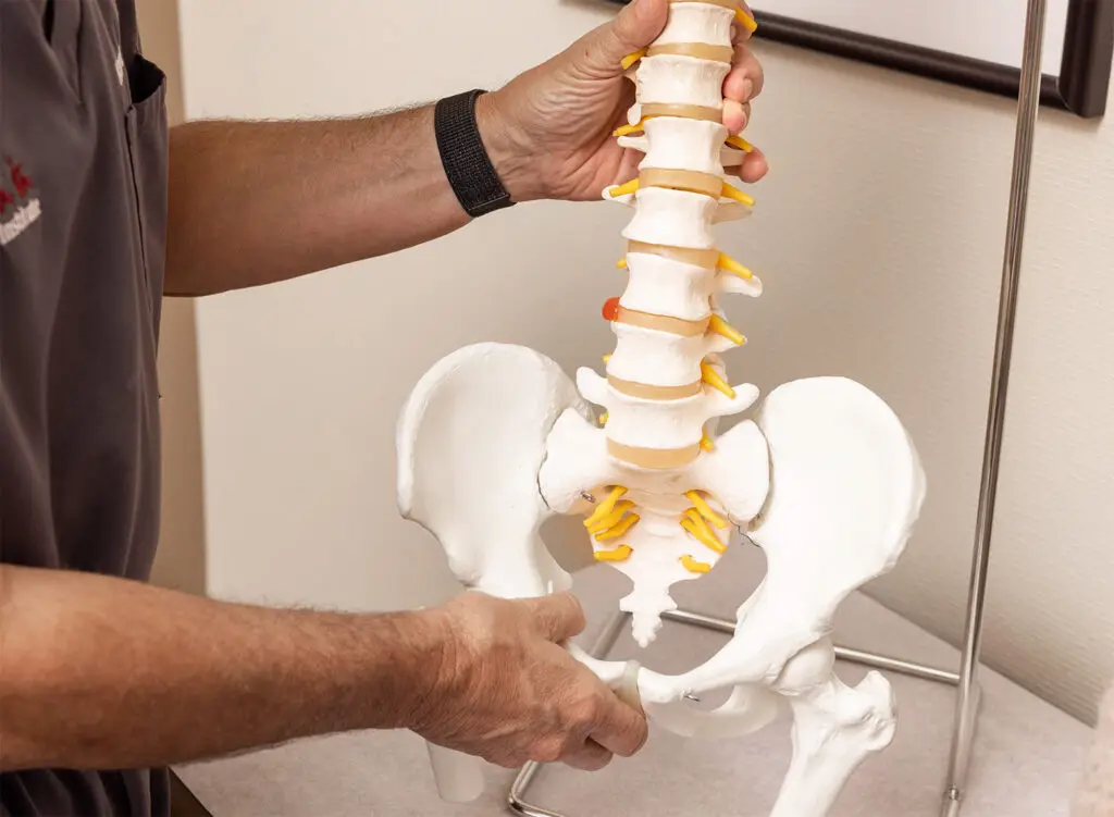
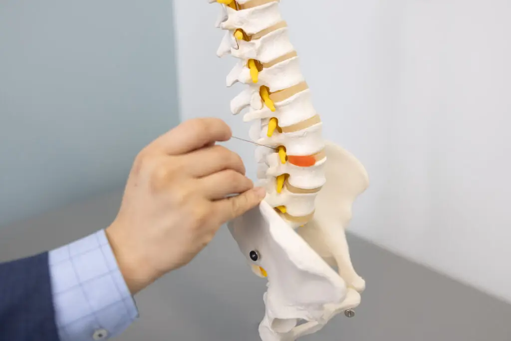
The Sacroiliac Joint (SI joint) is located between the sacrum (which supports the spine) and the ilium bones (which support the sacrum) of the pelvis. As the link between the ilium bones and the sacrum, the SI joint is responsible for transferring the weight of the upper body to the lower extremities.
Pain in this region arises from degenerative changes within the SI joint which may cause inflammation. Sacroiliitis is an inflammation of one or both immovable joints formed by the bones of the pelvis.
Patients with SI joint inflammation may have any or all the following symptoms:
- Lower back pain
- Sensation of lower extremity pain, numbness, tingling, weakness
- Pelvis / buttock pain
- Hip or groin pain
- Feeling of leg buckling or instability
- Disturbed sleep patterns due to pain
- Unable to sit for long periods
- Pain when going from sitting to standing
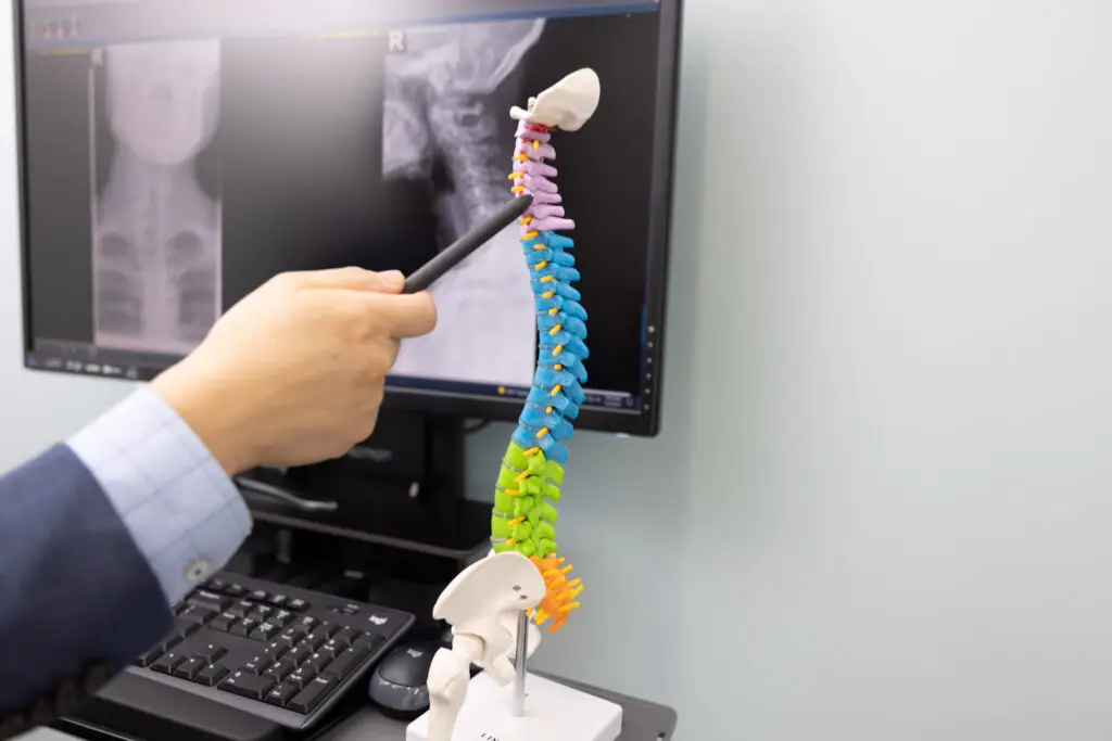
(insert photo of TBI doctor examining an x-ray)
Diagnosis involves physical examination, X-rays, MRI scans, and anesthetic injections into the SI joint as a diagnostic evaluation. If the pain decreases significantly after the injection, this helps to verify the SI joint as a source of pain. Injections may provide temporary pain relief, or the pain may remain reduced for an extended period. However, if the pain persists after conservative care treatment, a rhizotomy procedure may be necessary.
Patients Ask:
I have been diagnosed with facet joint syndrome. What happens next?
Texas Back Institute Responds:
Facet joint syndrome is most often found in the neck (cervical region) and lower back (lumbar) and is caused by age-related wear and tear on the joints over time. If conservative care treatments – physical therapy, massage, chiropractic care, and medications – fail to help to manage and control facet joint pain, spine specialists may recommend injections, ablations, or surgery.
What is the Next Step After a Medial Branch Block?
After a medial branch block, the next step may involve further treatment options like facet joint injections or rhizotomy. Rhizotomy is an umbrella term that involves ablation methods and is typically only considered after conservative treatments like physical therapy or nerve blocks have been explored.
Ablation methods used to disrupt problematic nerve signals include endoscopic surgery (link to minimally invasive surgery page) or radiofrequency ablation (RFA). Surgeons may decide to cut the nerves using open surgery or an endoscopic, minimally invasive approach, making a tiny incision with a camera instrument. RFA is performed using focalized high-intensity radio waves to cauterize the nerve.
Rhizotomies are performed under general or local anesthesia, depending on the location of the nerve. Surgeons perform this procedure using X-ray, fluoroscopy, or another image-guided technique to ensure precision.
- Endoscopic Rhizotomy: The surgeon uses a camera device called an endoscope to locate the affected nerve and sever its fibers. The endoscope is inserted through a small incision via a series of tubes called a tubular retractor system. This allows the surgeon to get to the nerve while bypassing healthy organs and tissues. This procedure is also called direct visualized rhizotomy.
- Radiofrequency Rhizotomy: Also known as radiofrequency ablation, a surgeon uses a radiofrequency current to ablate the problematic nerve fibers. RFA is commonly used to treat chronic pain conditions like osteoarthritis, facet joint syndrome, or SI joint pain.
Patients Ask:
How will I know if I am a good candidate for a rhizotomy?
Texas Back Institute Responds: The spine specialists at Texas Back will assess whether you are a good candidate for rhizotomy based on factors such as the severity of your pain, the location of affected nerves, and your overall health.
Recovery
The primary purpose of having a rhizotomy procedure is to alleviate pain. While some patients experience immediate relief, others may take up to two weeks to notice significant improvement. A complete recovery can take a few weeks. It is important to follow your doctor’s instructions regarding activity levels, pain management, and any physical therapy recommended.
A rhizotomy procedure is minimally invasive, takes less than an hour, and can be done on an outpatient basis. After the procedure, patients will typically spend thirty minutes to an hour in the recovery room. Patients should not drive or operate machinery for the next 24 hours. Patients should not engage in any rigorous activity for the first 24 hours. It is not uncommon to experience some pain, swelling and/or bruising at the surgical site. Depending on how well they feel, a patient may return to work one or two days after the procedure.
The duration of pain relief after a rhizotomy can vary from person to person. However, it typically lasts several months to a year. Some individuals experience longer-lasting relief, while others may need repeat procedures if the pain returns.

Patients Ask:
Will I need physical therapy after the rhizotomy procedure?
Texas Back Institute Responds: Physical therapy can be beneficial to help patients to regain strength, mobility, and flexibility to prevent muscle stiffness and improve overall function. If a patient is experiencing some residual pain after the rhizotomy procedure, physical therapists may use heat, ice, or ultrasound to alleviate discomfort.
Leading-Edge Technology Combined with Exemplary Patient Care
The rhizotomy procedure is one of many medical technological advances deployed by the world-class spine experts at Texas Back Institute. However, outstanding patient care is equally important for a patient’s recovery. Is back pain interfering with your quality of life? If the pain continues for more than a few weeks, contact the spine specialists at Texas Back Institute. Click here to schedule an appointment.
Learn more
Frequently Asked Questions
Medial branch blocks are commonly used to diagnose facet joint pain, while sacroiliac joint pain is typically diagnosed through a combination of clinical evaluation and medical imaging.
Facet joint syndrome is most often found in the neck (cervical region) and lower back (lumbar) and is caused by age-related wear and tear on the joints over time. If conservative care treatments – physical therapy, massage, chiropractic care, and medications – fail to help to manage and control facet joint pain, spine specialists may recommend injections, ablations, or surgery.
The spine specialists at Texas Back will assess whether you are a good candidate for rhizotomy based on factors such as the severity of your pain, the location of affected nerves, and your overall health.
Physical therapy can be beneficial to help patients to regain strength, mobility, and flexibility to prevent muscle stiffness and improve overall function. If a patient is experiencing some residual pain after the rhizotomy procedure, physical therapists may use heat, ice, or ultrasound to alleviate discomfort.
Locations


