Surgeons in every medical specialty understand the importance of precision in performing procedures. This is certainly the case with spine surgery.
Successfully correcting back and neck injuries, treating spinal disease or completely replacing parts of the spine with man-made devices demand extremely precise diagnosis and surgical procedures. The spine specialists at Texas Back Institute have always leveraged the latest technologies to attain the highest degree of precision possible.
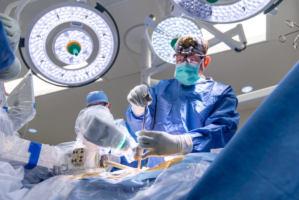
The diagnostic and surgical technologies for treating injuries and diseases of the spine are changing and improving exponentially over a shorter and shorter time frame. For more than 45 years, the world-class spine surgeons and clinicians at Texas Back Institute have understood the benefits of harnessing the latest technology such as 3D printing, robotics and imaging tools such as magnetic resonance (MRI) and computed tomography (CT) scans and using them to formulate state-of-the-art patient treatment.
In its storied history, Texas Back Institute has always been one of the leaders in medical technology and training. This commitment has enabled the organization to attract the “best and the brightest” surgeons and, most importantly, deliver exemplary patient care. If you are experiencing back or neck pain, contact us for an appointment. We will put that technology to work for you!
3D Printing is Changing Spine Surgery
While most of the mainstream and technology press coverage about 3D printing (3DP) concerns its use in the manufacturing of structures or other objects (everything from coffee cups to entire homes!), this technology can also increase the efficiency and success of other surgical challenges such as pre-operative planning, diagnosis and the surgical procedure itself.
Patient-specific 3DP models of anatomical structures can be generated from various medical imaging modalities in a multi-step process. In order to understand how spine surgeons use computed tomography (CT) and magnetic resonance imaging (MRI) volumetric datasets to improve surgical outcomes, we spoke with two spine experts – Dr. Peter Derman and Dr. Richard Guyer.

Dr. Peter Derman, Orthopedic Spine Surgeon at Texas Back Institute
“The data from CT scans and MRIs can be used to create virtual three-dimensional renderings of the spine, which can be extremely valuable,” Dr. Derman said. “Even the healthy spine exhibits complex geometry, but this complexity is magnified in the setting of spinal pathology (e.g., degeneration, scoliosis, trauma, infection, tumors).
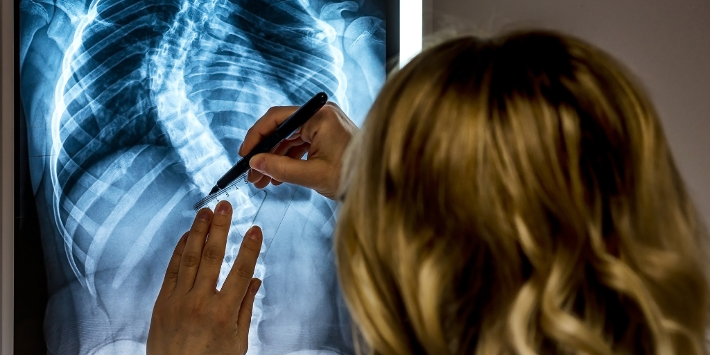
“It is extremely important to understand each patient’s anatomy when diagnosing a spinal issue and when planning and executing a surgical procedure. Surgeons use MRIs and CT scans before surgery to determine which techniques to employ and to help determine which implants they intend to use to achieve surgical goals. CT scans or other 3D imaging modalities can also be used in the operating room to help the surgeon insert these implants safely and to confirm that they have been placed correctly.”
Which Surgical Procedures Benefit From 3DP?

Dr. Richard Guyer, Orthopedic Spine Surgeon at Texas Back Institute
As one of the founders of Texas Back Institute, Dr. Richard Guyer has been a part of almost every technological advance in spine surgery for half a century. He offered his thoughts on the impact of 3DP.
“3D-enhanced imaging is most useful in diagnosing and treating complex deformity. It helps the surgeon understand what will be encountered in surgery.
“Due to the changes in the vertebral bodies and the connecting joints that occur when the spine begins to twist and turn in deformities, it is helpful for the surgeon to see these in a 3D printed model beforehand. With advancement in intraoperative navigation along with robotics, planning for successful correction is made easier for the surgeon.”
Dr. Derman adds, “Three-dimensional printing has been utilized in a variety of ways in the setting of spinal surgery. One use is to print 3D models of the spine before surgery so that the surgeon can manipulate and examine these before the procedure to get a better understanding of the issue. As Dr. Guyer said, this is most helpful when the spinal anatomy is particularly distorted by the issue (e.g., severe scoliosis).
“3DP can also be used to create custom forms that snap on to the patient’s vertebrae and act as guides for placing screws into the spine during surgery.
“Currently the most common use of 3D printing in spine surgery is to create interbody cages, which are spacers that are placed between the spinal bones to provide support and encourage bone growth during fusion operations. These cages are typically made of titanium, and the 3DP process allows for specific geometries to be created to optimize cage properties such as stiffness and the tendency to encourage bone growth.
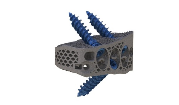
“Most of the 3D printed cages presently on the market are of standard sizes, but the next frontier is truly patient-specific implants. The data from a preoperative scan can be used to help design implants that more closely conform to an individual patient’s anatomy. These are then 3D printed, sterilized, and inserted during surgery, providing a tailor-made construct that I am optimistic will improve surgical outcomes such as better spinal correction, fewer fractures of the adjacent vertebral bones, and higher fusion rates.”
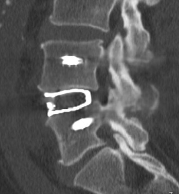
CT Scan of a patient with a 3D- printed patient specific interbody cage in the lumbar spine.
Artificial Discs and Other Surgical Applications for 3D Printing
According to Dr. Derman, “Three-dimensional printing is not yet being used in the setting of artificial disc replacements, although having patient-specific endplates on these devices would theoretically confer benefits. A custom device could better cover either side of the vertebral bodies, reducing the chance of fracture or undesired bone growth.”
Dr. Guyer adds, “Probably one of the best uses of 3DP implants is in spinal tumor resection and reconstruction. Surgeons are now able to take pre-op CT scans planning for excision of massive sacral tumors, for example, and then produce a near exact fitting 3DP implant from the post excision CT scan. The time for production of these implants has gone from over a week to days in many cases. In the future, they will be produced in the operating room.
“Another potential use of 3DP is in the production of patient specific implants in the lumbar spine, which exactly match the anatomy of the patient and allow for safer placement and hopefully faster fusion. This must be proven in future patient tests. Eventually we would like to have a patient specific artificial disc endplate for both the neck and low back. However, there will be FDA hurdles to navigate before this becomes standard practice.”
Other Promising Technologies on the Horizon
Dr. Guyer is optimistic that new technologies will continue to enhance positive patient care. “There are many advances that are making it easier and safer for the patients and the surgeon,” he said. “For example, we now have a technology that can convert MRI images into a CT- like images for planning proper screw placements. Since sometimes more than one CT scan is needed introspectively, this would eliminate the risk of radiation exposure to the patient.
“In the near future, multi-arm robotics will replace single-armed robotics in assisting the surgeon in placing hardware. As technology advances, we will see robotic assisted decompressions which are among the most common procedures in spine surgery. Finally, navigation along with AR (augmented reality) will allow more accurate placement of implants and artificial discs in the future.”
Dr. Derman is also excited about technological advances in spine surgery. He concluded, “We are in the midst of a revolution in spine care. A variety of ‘enabling technologies’ are being used to allow us to perform spinal surgeries more safely and less invasively. These include 3D navigation, robotics, augmented reality and endoscopic spine surgery. While helpful in their current form, these technologies will continue to improve over time, becoming even more powerful.
“I am particularly passionate about endoscopic surgery, in which a narrow camera is inserted through a dime-sized incision to ‘scope’ the spine, alleviating pressure from spinal nerves and addressing sciatica. Rather than a traditional procedure that requires opening the spine and damaging the surrounding muscles and ligaments, endoscopic techniques preserve these surrounding soft tissues and allow for more rapid recoveries.”
Texas Back Institute has been recognized as a leader in the use of minimally invasive surgery.
The latest technology combined with compassionate patient care is truly the best prescription and it is found at Texas Back Institute. If back pain is ruining your quality of life, we can help. Click here and make an appointment today.